


The desire to look ever more clearly and deeply into the human body has tantalised doctors for generations. Prof Terry Young FBCS and Dr Jonathan Nash FRCR, consultant breast radiologist, explore radiology’s past, present and future.
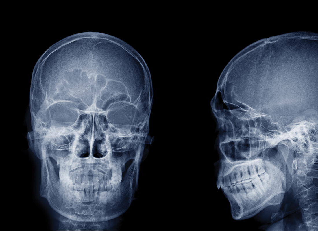
History shows how medicine is in constant flux. Many great minds have focused on staving off death’s inevitable call and improving the lot of the sick. For example, Wilhelm Roentgen’s work with X-rays in late 1895 triggered one of medicine’s most dramatic journeys: the birth of what we now call ‘clinical radiology’.
Since then, we have refined diagnostic imaging so that it is at the heart of many clinical pathways. New ways to probe the body and see inside are emerging all the time.
Turn back the clock
Since the 1980s, X-rays, other scans and diagnostic tests have been generating large datasets. Originally, images were captured in analogue form on film and paper. Such forms of information were neither easily reproduced nor transferred. Moreover, they were expensive to produce, using silver to form images in the case of film-screen combinations. These forms of information were all too easily lost and were susceptible to relatively rapid degradation. Indeed, the costs of production and storage combined with that short shelf-life became the principal determinants for retention strategies.
The seminal change was driven by the increasing digitisation of images, first at machine level, until those datasets drove powerful diagnostic tools in ways Roentgen could not possibly have imagined.
A step towards the future
As an exemplar in health, radiology has an enviable track record. The specialism has demonstrated an ability to develop, harness and enhance health informatics, and all associated IT, far ahead of other branches of medicine.
We’ll explore four aspects of this evolving story: the uptake of information systems to manage the work around images, image storage, image processing and automated diagnosis
Workflow
Radiology is a distributed service. Each phase – imaging, reading and consultation – takes place in different geographical locations. This provides an incentive to manage the end-to-end process.
Early digital solutions suffered by virtue of the limited technical state of the art. But, these systems are evolving rapidly to support clinicians and are placing the needs of patients – within whole healthcare systems – at their forefront. In turn, this drives not only how data is created, but how it is stored, transferred, interrogated and exploited.
Image storage
Today’s challenge is the volume of data that diagnostic services generate. Still-frame 2D images are ultra-high resolution and these often have multiple layers of information.
Beyond 2D, there are many different imagining systems which are capable of generating 3D and even 4D datasets.
Here, 4D describes multiple 3D images that are captured and presented over time. Antenatal 4D ultrasound scanning is perhaps the highest profile example of this, offering expectant parents the opportunity to see surface rendered 3D representation of the unborn child as a video clip.
The clinical appetite for these systems is proving insatiable because they offer such rich diagnostic information. Moreover, the more data that is available, the more demand there is to do more with it!
The lynchpin of radiology systems is the Picture Archiving and Communication System (PACS).
Image processing
Originally based around a local infrastructure from the 1990s, PACS now finds itself seated, in real time, within complex IT ecosystems that support cross-site and cross-enterprise data exchange. PACS’ rapid evolution has driven expectations to do more with the data. This, in turn, has driven standardised approaches to labelling the data, such as Digital Imaging and Communications in Medicine (DICOM), resilient high-speed communications and advanced processing and analytic capabilities.
To manipulate and present images, standard imaging dataflows enable standard mathematical algorithms to be applied. For example, we can create a dataset from a collection of photoelectric events, turn it into something manageable and of high diagnostic value, and finally present it on a display. We can go further by interrogating collections of datasets, looking for patterns and trends that deliver information far beyond what can be inferred from a single image. Even then, we are still some way from achieving the potential for population-wide benefit.
Automated diagnosis
In 1936, Alan Turning hypothesised devices that we now know as ‘Turing machines’, and went on to unpick the German Enigma cyphers and, ultimately, altered the course of the Second World War. From here, computers evolved still further and, in 1956, the concept of artificial intelligence was envisaged by John McCarthy at the Dartmouth Conference.
For you
Be part of something bigger, join BCS, The Chartered Institute for IT.
The data-rich nature of diagnostic imaging makes it an ideal target for health tech developers to apply the latest artificial intelligence approaches. And, they are doing so across the many steps in radiology workflows. These include not only interpreting images, but also identifying whom to image and when, and how to optimise the whole workflow to reduce burdens on high-cost specialist teams.
A clinical example: breast screening
The wealth of clinical evidence shows screening for breast cancer is best done with full-field digital mammography. It is a simple test to perform, with well-maintained modern systems using very small radiation exposures. The mammograms produced are packed with data that, even in their reduced ‘for presentation’ state, require the very highest resolution diagnostic displays for proper interpretation.
Most developed nations offer breast screening. These are offered to women between the ages of 40 and 75 and at intervals between one and three years. The programmes are run either locally or at a population-wide level.
Interpretation is undertaken by either one or two imaging specialists, with most women being reassured. A small proportion of women screened are recalled for further tests and a small proportion of those are diagnosed with breast cancer.
This process is always heavily scrutinised for quality and is necessarily laborious. The aim is to ensure cancer detection is high, without causing excessive levels of anxiety in patients or prompting additional tests that are not needed.
The high demand for limited specialist resources in breast screening combined with interruptions to services, for instance through the COVID pandemic, have left many services in dire straits. Backlogs are widespread and have increased screening intervals by 12 months across the UK and the Netherlands.
Backlogs and delays of this type result in missed opportunities for detecting cancers before they become symptomatic.
Population screening programmes generate huge amounts of data very quickly but are indifferent to individuals by design. The promise of artificial intelligence in this setting is therefore especially rich. It ranges from risk-stratifying women using historic data to processing multiple classes of information to predict breast cancer risk. Artificial intelligence could also automate image interpretation and guide treatment strategies that are most likely to be effective for individual women.
We have, for several years, been training artificial intelligence systems on data from human specialists’ performance.
These systems have been ingesting imaging and outcome data and producing ever improving results in automated image interpretation. The next step in that process will be setting artificial intelligence systems free to learn new lessons from the vast data lakes humans cannot hope to understand.
Summary
Radiology is a highly technically advanced medical speciality that generates very large datasets very quickly. Because of this, and in tandem with their evolution, it has innovated sophisticated systems to manage and learn from these datasets.
Legacies of analogue and human styles of working persist. But, real opportunities exist to harness the potential of digital data and to drive new ways of working locally, cross-enterprise and at population-wide levels.
Many challenges do, however, remain. While embracing the fierce and rapid pace of technical development, we must keep patients safe. Sound clinical governance is also important when adapting to changes. Finally, we must ensure that everybody in society – not just the digitally and socially advantaged – can benefit from these changes.
About the authors
After lengthy spells in research and development, and as a university professor in healthcare systems, Terry now runs Datchet Consulting which specialises in healthcare and innovation. Jonathan practised as a consultant breast radiologist with an interest in digital transformation in the UK for 11 years before shifting to industry, becoming medical director at Kheiron Medical Technologies last year.